Vitrectomy and vitreoretinal eye surgery

Vitreoretinal eye surgery includes a group of procedures performed deep inside the eye's interior with lasers or conventional surgical instruments.
As the name implies, this delicate surgery takes place on the gel-like vitreous and the light-sensitive membrane (retina) in the back of the eye.
Who performs vitreoretinal surgery?
If you need surgery on the vitreous or retina of the eye, your eye care professional will typically refer you to a specialist called a vitreoretinal surgeon.
This type of doctor specializes in the medical and surgical management of vitreoretinal disorders.
Conditions requiring a vitrectomy
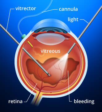
A vitrectomy surgery removes and replaces the vitreous humor or gel-like substance in the back of the eye. This procedure can address vision problems caused when foreign matter invades this usually pristine area of the eye's interior. One example of foreign matter is blood, from conditions such as diabetic retinopathy.
Light rays passing through the eye cause the foreign matter to cast shadows on the retina, resulting in distorted or greatly reduced vision.
The most common reasons for a vitrectomy include:
Diabetic retinopathy
Endophthalmitis (a serious infection inside the eye)
Sometimes, a vitrectomy also is used to treat large, persistent spots and floaters in the back of the eye.
The vitrectomy procedure
A vitrectomy often is performed under local anesthesia. However, general anesthesia is used in certain situations, especially when local anesthesia would be inappropriate, such as for people with inability to lie still.
Your surgeon will make three tiny incisions in the eye to create openings for the various instruments that will be inserted to complete the vitrectomy.
The instruments that pass through these incisions include:
Light pipe, which serves as a microscopic, high-intensity flashlight for use within the eye.
Infusion port, used to replace fluid in the eye with a saline solution and to maintain proper eye pressure.
Vitrector, or cutting device, that removes the eye's vitreous gel in a slow, controlled fashion. It protects the delicate retina by reducing traction while the vitreous humor is removed.
Once the surgeon removes the vitreous humor and clears the area, a saline liquid is used to replace the vitreous and maintain the proper shape of the eye and internal eye pressure.
What to expect after a vitrectomy
Because so many variables are involved, only your eye surgeon familiar with your condition can give you a realistic idea of what to expect following a vitrectomy.
But the underlying reason for the procedure usually is a major factor in determining how fast you will recover as well as the ultimate outcome.
After a procedure, you likely will use antibiotic eye drops for about the first week and anti-inflammatory eye drop medications for several weeks.
Follow your surgeon's advice carefully. In general, don't expect to know your final visual outcome for at least a few weeks. Again, your surgeon or attending eye care professional will be the best judge of your individual recovery.
Vitrectomies have a very high success rate. Bleeding, infection, progression of cataract and retinal detachment are potential problems, but these complications are relatively unusual.
For most patients who undergo a vitrectomy, sight is restored or significantly improved.
FIND AN OPTICIAN: An optician can assess your vision, find an optician near you.
Page published on Monday, 14 March 2022


