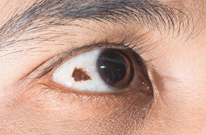
A closer look at the eye freckles on the whites of your eye

What is a conjunctival nevus?
A conjunctival nevus is a harmless accumulation of melanin-producing melanocyte cells on the conjunctiva, the clear tissue covering the eye. Melanin is the substance that gives our skin and hair its color. A conjunctival nevus can be present when you are born or develop over time.
A conjunctival nevus may also be referred to as a benign melanocytic tumor or lesion.
What does a conjunctival nevus look like?
The appearance of a conjunctival nevus (plural nevi) is dependent on the individual accumulation of melanin pigment. It can vary in appearance from dark brown to light brown to yellow — or somewhere in between. About half of conjunctival nevi also have a clear cyst associated with them.
SEE RELATED: Linear Nevus Sebaceous Syndrome
Does a conjunctival nevus have any symptoms?
A conjunctival nevus does not typically cause any symptoms. It also does not usually affect vision. This is because the nevus is not located in the direct line of sight where light rays enter the pupil of the eye.
What can cause a conjunctival nevus?
Conjunctival nevi are colored spots that are the result of an accumulation of melanocyte cells on the conjunctiva. These cells are responsible for producing melanin pigment. Instead of spreading out uniformly, they have clumped together on the eye’s conjunctiva.
People are usually either born with this nevus or develop it in their first two decades of life.
Can a conjunctival nevus change over time?
Yes, a conjunctival nevus can change over time. These eye freckles can darken or lighten with age.
According to a study published in JAMA Ophthalmology of more than 400 people, color change was detected in 13%. And a change in size was detected in 8%.
A conjunctival nevus is similar to a freckle or mole on the skin. Hormone shifts, such as those caused by puberty or pregnancy, may result in color and size changes. Oral contraceptives are also associated with an increase in nevus size.
There seems to be a link between exposure to ultraviolet rays and nevi. UV radiation may result in the development of, or a change in, the appearance of a nevus.
This is another reason that protecting your eyes by wearing sunglasses is good for your long-term eye health.
How is a conjunctival nevus diagnosed?
During a comprehensive eye exam, an eye doctor can diagnose a conjunctival nevus. Eye doctors use an instrument called a slit lamp, which allows them to examine the front of the eye. This gives them the ability to view the nevus with a bright light and under high magnification.
An eye doctor will make a note of the appearance and size of the nevus in your medical record. They may even take a picture. This gives them the ability to monitor any changes in the appearance of the nevus at your next exam.
Are there other types of colored spots on the conjunctiva?
In addition to nevi, there can be other types of pigmentation located on the conjunctiva. This includes:
Racial melanosis — benign, flat, pigmented areas in both eyes. It is seen in people with darker colored skin.
Primary acquired melanosis (PAM) — benign, flat, pigmented area found in one eye, typically in Caucasians. According to some studies, three-fourths of conjunctival melanomas (cancer) are from PAM.
Conjunctival melanoma— cancer of the melanocyte cells on the conjunctiva. These will require surgical removal and follow-up treatment depending on the melanoma.
Is a conjunctival nevus dangerous?
Conjunctival nevi are benign, meaning they are not harmful. A conjunctival nevus that is present at birth has a low risk of becoming cancerous. A freckle that develops later in life has a slightly higher chance of becoming cancerous, although the risk is still low.
The risk of a conjunctival nevus developing into a conjunctival melanoma is less than 1% over someone’s life.
If a conjunctival nevus appears to be growing larger or changing in appearance, it is important to see an eye doctor so that they can evaluate it. Although it is not common, a conjunctival nevus can develop into conjunctival melanoma.
How is a conjunctival nevus treated?
Since conjunctival nevi are typically not dangerous and have no symptoms, they do not usually need treatment or removal. Most often, an eye doctor will recommend monitoring the nevus.
If an eye doctor finds that a nevus has become cancerous and developed into a conjunctival melanoma, they will refer management and treatment to a specialist. Depending on how advanced the melanoma is, treatment may include:
Excision or surgical removal
Radiation
Laser therapy
Enucleation (removal of the eye) in very rare cases
Should I see a doctor for a conjunctival nevus?
Yes. Although a conjunctival nevus is a benign condition, it is important for an eye doctor to monitor it. This is to ensure that there are no changes that could indicate the development of melanoma.
It is important to be seen by an eye doctor if you notice:
Change in appearance of the conjunctival nevus
Increase in size of the conjunctival nevus
If no changes are noticed in the appearance, a conjunctival nevus can be monitored as part of a routine, comprehensive eye exam.
READ NEXT: Choroidal nevus
Nevus (eye freckle). American Academy of Ophthalmology. January 2022.
Conjunctival melanocytic tumors. EyeWiki. American Academy of Ophthalmology. February 2022.
Conjunctival nevus. Wills Eye Hospital. Accessed April 2022.
Answer: Can you identify this condition? Canadian Family Physician. October 2011.
Conjunctival nevi: Clinical features and natural course in 410 consecutive patients. Archives of Ophthalmology. February 2004.
Conjunctival nevi. Columbia University Department of Ophthalmology. Accessed April 2022.
Page published on Wednesday, June 8, 2022


