What to expect during a comprehensive eye exam

Schedule an exam
Find Eye DoctorWhat is a comprehensive eye exam?
A comprehensive eye exam is a full checkup for your eyes. It consists of several noninvasive eye tests so your eye doctor can assess your total eye health, not just your vision. Like yearly checkups with a family doctor, it’s very important to have routine eye exams with an optometrist.
Many eye conditions don't cause any symptoms or noticeable vision changes. And, unfortunately, vision screenings and basic vision tests can often miss these problems. This is why having yearly comprehensive eye exams is the best way to stay on top of your overall eye health. They allow you and your eye doctor to catch any issues before they can lead to vision loss.
Plus, a comprehensive eye exam could even alert you to other types of health concerns. Routine eye exams can detect the early signs of over 250 different systemic conditions. Many people are able to take important preventive steps for their health due to early warnings from their eye exams.
What's the difference between an eye test and an eye exam?
An easy way to remember the difference is that an exam usually includes more than one eye test. A comprehensive eye exam includes several eye tests, procedures and even other exams.
However, it's fairly common for people to use eye exam and eye test interchangeably. Depending on where you live, optometrists might refer to an eye exam as a comprehensive eye test.
Basic vision tests might also be called eye exams in some cases. For example, some optical stores might offer online "eye exams." These online eye tests consist of digital vision and refraction tests to find your lens prescription. They are not comprehensive eye examinations.
Preparing for a comprehensive eye exam
If it's been a while since your last full eye exam, there are a few things you can do to help ensure a seamless visit. For example, you may want to make a list of things you need to bring, as well as any questions you have.
Visiting your eye doctor is a lot like visiting your general physician. They'll ask you for a lot of the same types of information, so having a list of what to bring to your eye exam can be really helpful. For example, it may help to bring:
The names and dosages of any current medications (including eye drops), supplements, etc.
Your personal and family medical history
Your current glasses and lens prescription
Your insurance card and information
A list of any vision changes or symptoms you've noticed since your last exam
Having all of this prepared is especially helpful if you're seeing a new eye doctor for the first time. But even if you've seen them in the past, they'll need to update their records with any changes since your last visit.
Writing down any questions you have can help you make sure nothing slips your mind before or during your visit. Usually, you'll be given any information you need when you book your eye exam. But some of the questions you may want to ask your eye doctor or their staff ahead of time include:
If you need to arrive earlier than your appointment time
How long the exam usually lasts
Whether your eye exam will include pupil dilation (if yes, you may need to have someone that can drive you home)
Whether they accept your vision and/or medical insurance
Whether there may be any charges not covered by insurance
If you don't have vision or medical coverage, how much their exam typically costs
If they need you to bring anything not already on your list
Eye symptoms, past eye exams and eye injuries
If you wear glasses or contact lenses, you should bring them to your exam. And if you’re seeing the eye doctor as a new patient, it’s also good to bring a copy of your written prescription if you have it.
They’ll also need to know about any eye symptoms or vision changes you've noticed. Try not to leave anything out, even if it seems minor or doesn't happen very often. A lot of eye care professionals use the following "FOLDAR" list when they ask about symptoms:
Frequency – How often do they happen?
Onset – How long ago did they begin?
Location – Which eye has the symptoms? Where on the eye?
Duration – How long do the symptoms usually last each time?
Associations – Have you noticed any times or situations when they seem better or worse?
Remedies – Have you found anything that improves the symptoms?
Eye injuries and surgeries are also important parts of your health history. Both have the potential to lead to problems later on.
Personal and family eye and vision health
If you've never had a comprehensive eye exam, you may not have a lot of personal eye health history to share. However, knowing about your family's eye health can be just as helpful for your eye doctor.
Tell them about any eye conditions you know your family members have had, such as:
Glaucoma
Macular degeneration
Cataracts
If you have had full exams before, it can be helpful to share information from those past eye exams with your new eye doctor. It allows them to see any changes in your eyes or vision that have happened since then. However, sharing those records isn’t required, and it can often be a big hassle to request them.
Either way, be sure to give the doctor a thorough history of your eye health, including:
Existing eye conditions
Previous diagnoses you may have had
Past or recurrent eye infections
Your history of eye surgery
Any signs or symptoms your previous eye doctor was monitoring
Your health history and current medications
Providing a thorough general health history to your eye doctor is a lot more important than you might think. Your eyes are very much a part of your overall health. This means eye doctors need to know about things that may seem unrelated to your eyes.
When you fill out your health history form, you should be just as detailed as you would be for your primary physician. Some things that may seem less obvious to tell your eye doctor about include:
Your current and past prescription medications
Any over-the-counter drugs, vitamins and supplements you use
Any recent illnesses or infections, even if they seemed mild or embarrassing
Your personal and family history of high blood pressure, diabetes, thyroid and autoimmune disorders, and other systemic health issues
Past cosmetic surgery and procedures, such as eyelid lifts, Botox and fillers
All these things can be relevant, so your eye doctor needs to know about them to fully assess your eye and vision health.
Eye exam cost and whether you have vision insurance
Understanding how much you might need to pay and how your insurance will work are also good ways to prepare.
The cost of a comprehensive eye exam can range a lot, from as low as $10 to as high as $200 or more. But the amount you pay will depend on your insurance coverage and several other factors, including:
Vision insurance – Having vision coverage can often keep your out-of-pocket costs at $40 or less. Without it, the average cost for comprehensive eye examinations is about $140.
Large chains vs. independent optometrists – Exams with independent eye doctors usually cost more than with big retailers. They can offer more in-depth exams and expertise than most chains.
Location – Full eye exams usually cost less in rural areas than in larger cities.
New vs. returning patients – Your first comprehensive eye exam as a new patient may cost a little more than your following full exams with that doctor.
Refraction tests – A refraction (the test that determines your lens prescription) is usually a standard part of a full eye exam — but not always. Make sure your exam includes a refraction test if you need an updated vision prescription for your glasses.
Additional tests – If you need any other eye tests that aren’t included in your comprehensive exam, these can add to your total cost.
Contact lens fittings – Fittings are not usually included in comprehensive eye exams. They’re a separate exam and aren’t always covered by vision insurance.
Lenses and frames – Contact lenses and eyeglasses are often totally separate from the cost of the eye exam. However, some large chains do offer bundles or reduced exam prices when you buy your glasses from their store.
What to expect when you arrive at your eye examination
When you get to your appointment, make sure to check in at the front desk before finding a seat. It’s best to let them know as soon as you’ve arrived.
Many practices will ask for your insurance or other payment information upfront. If you haven’t already, this is a good time to ask about your out-of-pocket costs for the exam.
You may also need to fill out some patient forms if you haven’t done them through an online patient portal. Even if you’ve already filled them out online, they may ask you to review them to make sure everything is correct.
What to expect during the eye exam procedure
Comprehensive eye exams are simple, pain-free processes. They can last from 30 to 90 minutes, but most of the eye test procedures only take a few minutes each.
The exact tests included can vary a little depending on the doctor, your eye health and your exam history. For example, if you have yearly comprehensive eye checkups, you may not need to have certain tests each time. But your first (or first in a long time) complete eye exam may include more tests than a routine eye checkup.
When it's time for your exam, someone will show you into an exam room. You'll have a seat in the exam chair, and the eye doctor or their assistant will introduce themselves and get started.
During the exam, they’ll ask you further questions about your health history. They may also talk with you about your job, daily activities and general lifestyle. This is so they can get a better understanding of anything that may be affecting your eyes and vision.
They'll explain each step along the way and will be happy to answer any questions you have. Even so, it can be helpful to know what to expect in the eye tests and procedures that happen during an eye exam.
Visual acuity test

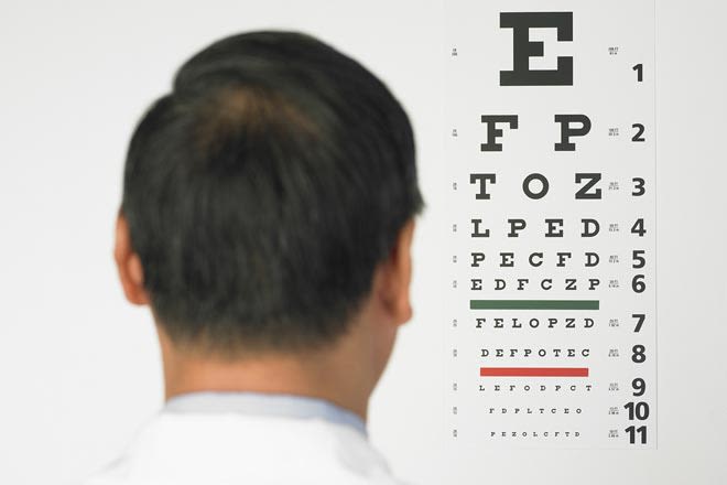
A standard eye chart.
Visual acuity is the measurement of the sharpness of your vision. One of the most common types of visual acuity test is done using the Snellen eye chart. It has several rows of letters, often with a large capital E at the top, that gradually get smaller.
The eye doctor can determine your visual acuity based on the smallest row you can see clearly from 20 feet away. This is where measurements such as 20/15, 20/20 and 20/40 come from.
You may also have a test for your near vision. This test is very similar, but the chart is on a smaller card that you'll hold to look at from about 14 inches away.
LEARN MORE: What is the Snellen eye chart?
Refraction test
If you need glasses, they will use a refraction test to find your vision prescription.
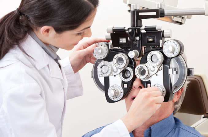
You'll look at an object (often the eye chart from the acuity test) through an instrument called a phoropter. Then the doctor will click through a series of lenses inside the phoropter and ask you which ones make the chart look the sharpest. This allows them to zero in on the lens prescription that brings you closest to 20/20 vision.
In some cases, they may do an autorefractor test before using the phoropter. An autorefractor is a device that shines light into your eye to calculate how the light hits your retina. This measurement can provide a good starting point for the rest of the manual refraction test.
Pupillary response test
This test evaluates how well your pupils react to light. The eye doctor will shine a small, bright light into each eye, remove it for a few seconds, and then do it again. This allows them to see how quickly and how much your pupils can contract and relax.
Ocular motility tests
These tests let the doctor assess your eye muscle function and eye alignment.
They will ask you to hold your head still and follow the movement of a pen or penlight with just your eyes. By watching your eyes move, they can look for signs of muscle weakness or misalignment that can affect how well your eyes work together as a team.
Each eye sends separate information to the brain, and then the brain combines it to create one clear, 3D image. This is called binocular vision. Your eye muscles need to keep your eyes perfectly aligned to give you binocular vision. When the eyes don't align correctly, this is a condition called strabismus.

Cover test to check eye alignment.
Another kind of test they may use to check for strabismus is called a cover test.
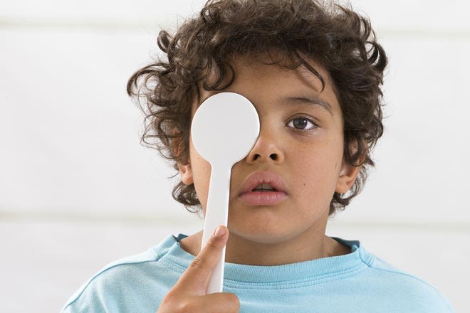
For this test, you'll stare straight ahead at one spot instead of following a penlight. They will cover each eye for a few seconds, one at a time. While each eye is covered, they'll watch for any shifts in the gaze direction of the other eye.
They might also repeat the process to perform a "cover/uncover" test. This time, they'll watch for gaze shifts in the covered eye as they are removing the cover.
Depth perception test
Depth perception is another important aspect of binocular vision. Your eyes need to be able to work in unison so you can judge distances and see in three dimensions.
There are several ways to test depth perception. Common methods involve looking at or touching special images while wearing polarized "3D" glasses. Some doctors may use a viewing device that shows each eye a different image at the same time.
They can assess your depth perception based on how you describe or select certain images.
Visual field test
This test evaluates your peripheral or "side" vision. In one common method, you'll look straight ahead with one eye covered. The doctor will hold an object or their fingers in the outer areas of your vision and move them around until you can see them.
They may also ask you to look straight ahead into a bowl-shaped device. It will display points of light or other images in your side vision, and you'll press a button when you see them.
Visual field tests can tell the doctor if you have any blind spots in your peripheral vision. Peripheral vision issues can be a sign of glaucoma, diabetic eye disease or other serious conditions.
Depending on any symptoms or previous diagnoses you have, your doctor may use more detailed test methods. They may also use an Amsler grid test to check your central vision. Issues with central vision can be a sign of macular degeneration or other problems in the macula.
Color blindness test
This test checks for color vision deficiencies, such as problems seeing red and green. A color vision deficiency may also cause less noticeable symptoms. For example, certain colors might look dull, or it might be hard to distinguish between shades of the same color.
Color vision tests are commonly called color blindness tests. They are usually done with images made up of colored dots. Some of the dots form a shape in the center of the image, and the rest act as background. A color vision deficiency can make it hard to see a center shape made of certain colors.
Color blindness tests are important for adults as well as for kids. People who are born with a color deficiency usually don't notice it because it's always been there. But people who develop a deficiency later on may not notice symptoms either. A later onset can be a sign of an eye or brain injury or other underlying conditions.
Tonometry
This test measures the pressure inside your eyes. It's a common part of full eye exams because high eye pressure is a serious risk factor for glaucoma. Glaucoma almost never has any symptoms, so eye pressure tests are key to catching it early.
There are a few different ways to test eye pressure, but the most common is applanation tonometry. For this method, they numb your eye first. Then the eye doctor gently presses a small device called a tonometer against your cornea.
They may also use a non-contact method called the "air puff in eye test" or "puff of air eye test.” This eye test isn't as accurate, so it's mostly used for screenings or for young children.
Corneal mapping
Corneal mapping is another name for corneal topography. It’s an imaging technique that creates a 3D map of the surface of your cornea. Corneal mapping isn't a typical part of everyone’s eye exam, but it may be included for patients with:
Certain contact lens fittings
Astigmatism
Keratoconus
Corneal growths
Corneal scarring from infections or injuries
Pre-eye surgery exams
You'll sit a few inches in front of the imaging device with your face supported by forehead and chin rests. As you look straight ahead, it will scan your eyes for a few seconds to take the images.
Pupil dilation
Dilating your pupils means making them bigger. For the dilated part of the exam, they'll give you special eye drops that temporarily expand your pupils and hold them open. This is an important step that allows a much wider view of the inside and back of your eyes. It also ensures that your pupils won't shrink during the exam.
Dilating eye drops start working in about 20 to 30 minutes. Most people will have blurry vision, trouble focusing and extra light sensitivity until the drops wear off. Their effects can last for several hours, so it's best to plan the rest of your day accordingly.
In this video, an eye doctor explains the importance of dilated eye exams. (Video: National Eye Institute)
Slit lamp examination
An eye doctor uses a slit lamp to examine all the tissues and structures of your eyes. It's a special type of microscope with bright lights that allows them to look very closely for any issues.
During a slit lamp exam, they will start by looking at:
The eyelids, eyelashes and other external tissues
The white of the eye, conjunctiva and cornea
The area between the cornea and the lens
The iris, pupil and lens
They'll move the slit lamp in front of your exam chair and have you rest your forehead and chin against it. They can then look at your eyes through the other side of the microscope.
After examining the front of the eye, the doctor will begin the retinal exam. They can do the retinal exam with the slit lamp by adding another small lens. They'll hold the lens very close to your eye and then use the slit lamp microscope to look through it into the back of your eye.
Retinal examination
Eye doctors look at the retina during an exam of the fundus, or back portion of the eye. Other names you may hear for this exam are fundoscopy and ophthalmoscopy.
The fundus contains the retina, optic nerve, important blood vessels and other structures that are vital to your eye health and vision. Fundus exams are key in detecting and monitoring serious eye conditions such as:
Age-related macular degeneration
Glaucoma
Blood vessel damage due to hypertension or diabetes
A torn or detached retina
There are a few different ways to examine the fundus. One common method is to use the slit lamp. In another method, called direct ophthalmoscopy, they’ll use an ophthalmoscope. This is a small, handheld device with a bright light and special magnifying lenses.
Indirect binocular ophthalmoscopy is also common. For this method, they wear special binoculars on a headband with a very bright light to view the back of the eyes.
Each method allows a slightly different view of the fundus, and some may not require dilation. Your eye doctor will determine which is best for you based on your health history and any symptoms you may have.
They may also recommend further tests if they see any signs of problems. This could include retinal imaging, such as optical coherence tomography (OCT), or other imaging techniques.
LEARN MORE: What is Optomap retinal imaging?
What to expect after the eye exam
When the eye exam is complete, your doctor will go over all the results with you and answer any questions you have. You'll also receive a copy of your lens prescription if you need vision correction.
Depending on your results, they may need to recommend further eye tests. Often, you can have these tests at follow-up visits with the same doctor, or they may refer you to a specialist.
They'll also discuss treatment options with you if they've made a new diagnosis. If you have an existing diagnosis, they'll review your current treatment plan with you as well. This will include how often you should have comprehensive eye exams — yearly or more frequently — based on your results.
When you check out at the front desk, you’ll schedule your next visit or any follow-up eye tests you may need.
Eye exam. Cleveland Clinic. August 2024.
See the full picture of your health with an annual comprehensive eye exam. American Optometric Association. Accessed November 2024.
Comprehensive eye exams. American Optometric Association. Accessed November 2024.
The eye exam. Canadian Association of Optometrists. March 2023.
Comprehensive adult eye and vision examination. Second edition. American Optometric Association. January 2023.
Eye exam costs and what affects it. VisionCenter.org. September 2024.
What should I bring to my eye exam? Canadian Association of Optometrists. March 2023.
Comprehensive eye exam. Dean McGee Eye Institute. OU College of Medicine, Department of Ophthalmology. Accessed November 2024.
Taking patient history (for ophthalmic techs). Eyes On Eyecare. December 2019.
Ask your family about their history of eye disease. EyeSmart. American Academy of Ophthalmology. November 2017.
How to take a complete eye history. Community Eye Health Journal. December 2019.
Developing a constructive approach to case history. Review of Optometry. February 2023.
Comprehensive eye examination: What does it mean? Community Eye Health Journal. December 2019.
Eye exam cost without insurance: How much is it? Aflac. Accessed November 2024.
What you need to know: Eye exam costs and financing options. VisionCenter.org. September 2024.
Buying prescription glasses or contact lenses: Your rights. Federal Trade Commission Consumer Advice. June 2024.
How much do contact lenses & fittings cost? VisionCenter.org. September 2024.
Eye exams. Herbert Wertheim School of Optometry & Vision Science. UC Berkeley. Accessed November 2024.
Eye exam and vision testing basics. EyeSmart. American Academy of Ophthalmology. February 2024.
Standard eye exam. A.D.A.M. Medical Encyclopedia [Internet]. February 2023.
Visual acuity test. A.D.A.M. Medical Encyclopedia [Internet]. February 2023.
What is astigmatism? Symptoms, causes, diagnosis, treatment. EyeSmart. American Academy of Ophthalmology. October 2024.
Pupillary light reflex. StatPearls [Internet]. July 2023.
Extraocular muscle function testing. A.D.A.M. Medical Encyclopedia [Internet]. February 2023.
Binocular vision dysfunction (BVD). Cleveland Clinic. February 2024.
What is adult strabismus? EyeSmart. American Academy of Ophthalmology. September 2024.
Preliminary examination. In The Ophthalmic Assistant (Tenth Edition). 2018.
Depth perception test. Cleveland Clinic. January 2024.
Visual field test. Cleveland Clinic. April 2023.
Visual field test. EyeSmart. American Academy of Ophthalmology. March 2022.
Amsler grid eye test. Cleveland Clinic. November 2023.
Color blindness. National Eye Institute. August 2024.
Testing for color vision deficiency. National Eye Institute. August 2023.
Tonometry. StatPearls [Internet]. December 2023.
Eye pressure testing. EyeSmart. American Academy of Ophthalmology. April 2022.
Tonometry. Health Library. University of Michigan Health, Michigan Medicine. June 2023.
Corneal topography. EyeSmart. American Academy of Ophthalmology. May 2021.
What are dilating eye drops? EyeSmart. American Academy of Ophthalmology. October 2024.
What is a slit lamp? EyeSmart. American Academy of Ophthalmology. April 2018.
Slit lamp examination. EyeWiki. American Academy of Ophthalmology. May 2024.
Ophthalmoscopy. A.D.A.M. Medical Encyclopedia [Internet]. February 2023.
Ophthalmoscopy. MyHealth.Alberta.ca. June 2023.
Binocular indirect ophthalmoscopy. EyeWiki. American Academy of Ophthalmology. August 2024.
Retinal imaging. Cleveland Clinic. June 2023.
Page published on Wednesday, February 27, 2019
Page updated on Tuesday, November 26, 2024
Medically reviewed on Friday, November 15, 2024


