Epiblepharon

What is epiblepharon?
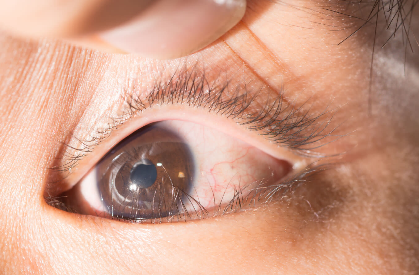
Epiblepharon is a congenital anomaly in which a loose fold of skin and the underlying muscle push the eyelashes upward and inward. The involved lid muscle is called the orbicularis oculi. It surrounds the eye and is used to close the eyelids. The resulting epiblepharon is more common in the lower than the upper eyelid.
When epiblepharon occurs, the lid folds over the eyelid margin where the skin and the surface of the eye meet. This causes the eyelashes to point vertically upward or inward, often rubbing and irritating the front surface of the eye. Importantly, epiblepharon does not typically impact vision.
Epiblepharon is most often congenital (present at birth), but it can also be acquired by patients with orbital (eye socket) conditions. Epiblepharon appears most commonly in East Asian populations.
Symptoms
Epiblepharon is often asymptomatic. But when people do have symptoms, they may be significant. Most symptoms are a result of the disruption to the cornea (the clear front surface of the eye).
The severity of symptoms themselves may depend on the number and thickness of the eyelashes that are rubbing against the cornea. A greater number of thick lashes will cause more significant symptoms than a small number of thin lashes. As lashes rub against the eye with each blink, the front surface of the cornea is disrupted.
The symptoms associated with epiblepharon include:
Foreign body sensation (the feeling that something is in your eye)
Excessive blinking
If epiblepharon isn’t properly managed or treated, a person’s eyelashes may rub against their eye enough to cause some of the following potentially serious complications:
Superficial punctate keratitis (a type of keratopathy)
Corneal infections
Corneal scarring or thinning
Induced astigmatism
Decreased vision due to scarring
READ MORE: Eye infections: Causes, symptoms and treatment
Causes
Congenital and acquired epiblepharon each have different sets of causes.
Congenital epiblepharon is the absent or weak attachment of the lower and upper eyelid retractors to the overlying skin. Some researchers have proposed that abnormal eyelid retractor development may be the cause.
Others believe it may have to do with a weak attachment of the eyelid’s orbicularis oculi muscle and skin to the tarsus, the connective tissue that gives the eyelids their shape.
While rare, acquired epiblepharon may occur due to secondary increases in intraocular pressure (IOP). Acquired epiblepharon may be associated with:
Orbital tumors
Obesity
Changes to the orbit or lower eyelid from surgery or trauma
Steroid use
Treatment
Congenital epiblepharon is primarily a pediatric condition. For many patients, the condition will resolve on its own as the eyelid and facial bones grow and develop. Management may include topical lubricants and proper follow-up until the condition resolves. If the epiblepharon persists, the need for additional treatment will depend on its clinical features.
Epiblepharon can be managed non-surgically unless significant complications are present. Non-surgical treatments include:
Lubricating artificial tears – These can be used to soothe irritation caused by the eyelashes rubbing against the cornea.
Hyaluronic acid gel injections – The goal of these injections is to turn the eyelashes back outward so they avoid contact with the cornea. This is a minimally invasive procedure and the effect typically lasts for around 19 months.
Botulinum toxin type A (BoNT-A) injections – BoNT-A injections are a safe and effective option for correcting symptomatic epiblepharon in patients younger than 2 years. Effects typically last up to six months.
Surgery may be necessary if symptoms are severe and/or persistent, or if corneal complications occur. The goal of surgically treating epiblepharon is to rotate the lashes outward to avoid ocular surface touch. Surgical options include:
Eyelid everting sutures – This procedure does not require an incision and is done in-office. It uses sutures to fold the lower eyelid retractors downward and reverse the upward migration of the lid and lashes.
Orbicularis oculi muscle and skin excision – This treatment tightens the orbicularis oculi muscle and removes excess skin from the eyelid. Scarring of the lid may result.
Transconjunctival approach – Unlike other surgical treatments for epiblepharon, this procedure for correcting lower eyelid retraction avoids external incisions and leaves very little risk for scarring.
In cases of acquired epiblepharon, treatment is usually targeted at the root condition. Addressing the underlying condition often reverses the epiblepharon and its clinical impact.
Prognosis
While epiblepharon can lead to potentially serious complications, the condition itself is not inherently dangerous or immediately threatening. Once it has been corrected, either naturally or with medical intervention, a patient's prognosis is usually quite good.
That said, untreated epiblepharon can cause severe symptoms and complications. If you suspect you or someone you know has epiblepharon, make an appointment with your eye doctor for proper evaluation.
READ NEXT: Treating your infant’s or child’s red or bloodshot eyes
Relationship between lower eyelid epiblepharon and epicanthus in Korean children. PLOS ONE. November 2017.
Congenital and acquired epiblepharon. EyeWiki (American Academy of Ophthalmology). August 2022.
Epiblepharon. American Association for Pediatric Ophthalmology & Strabismus. November 2021.
Management of epiblepharon state of the art. Current Opinion in Ophthalmology. September 2016.
Nonsurgical correction of epiblepharon using hyaluronic acid gel. Journal of American Association for Pediatric Ophthalmology and Strabismus. April 2018.
Treatment of symptomatic epiblepharon with Botulinum toxin type A in patients under 2 years of age. Archivos de la Sociedad Española de Oftalmología. January 2020.
Eyelid: Transverse everting sutures (Quickert sutures, three-suture technique). Operative Dictations in Ophthalmology. April 2017.
Pretarsal orbicularis oculi muscle tightening with skin flap excision in the treatment of lower eyelid involutional entropion. BMC Ophthalmology. December 2021.
Orbital decompression. Kellogg Eye Center. April 2015.
Page published on Wednesday, March 29, 2023
Page updated on Tuesday, April 4, 2023
Medically reviewed on Wednesday, December 21, 2022


