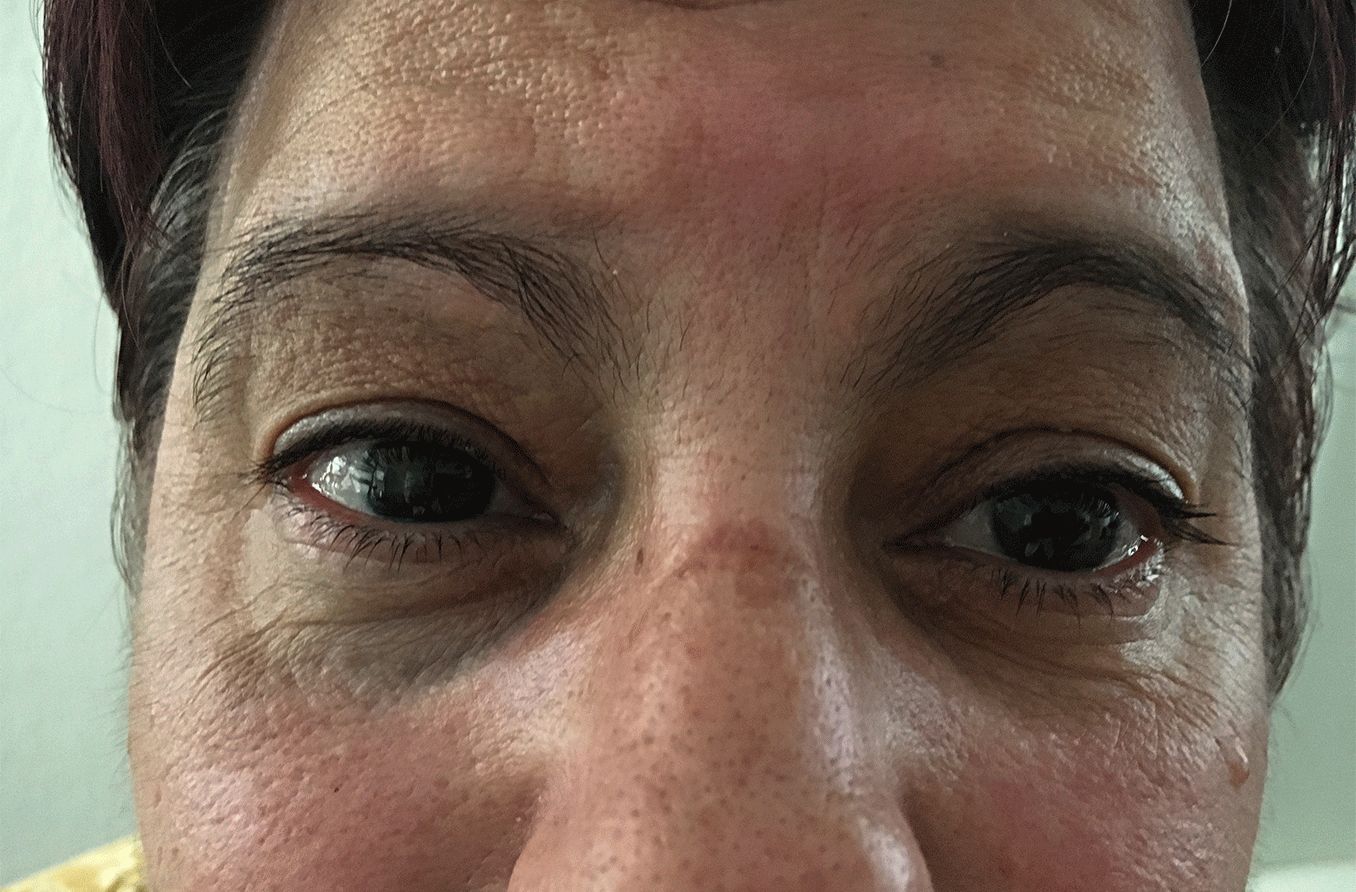
What is nevus of Ota?

Nevus of Ota
Nevus of Ota, also called oculodermal melanocytosis, is characterized by dark spots (called hyperpigmented lesions) on the face and one eye. It is a benign (non-cancerous) form of melanosis, which is a condition caused by an abnormal amount of melanin or other pigment-discoloring tissue.
The hyperpigmentation of a nevus of Ota lesion occurs due to melanocytes (pigment cells) becoming trapped deep in the tissue. When trapped, their color is affected.
Around 50% of people affected by nevus of Ota are born with it, while the rest develop it during their teenage years or adulthood.
Other conditions that feature hyperpigmented lesions are nevus of Ito and nevus of Hori. In nevus of Ito, lesions are present on the shoulder and upper arm. Nevus of Hori is distinguished by symmetric lesions most often located around the cheeks, nose and sides of the forehead.
Symptoms
Other than the presence of hyperpigmented lesions, nevus of Ota is usually asymptomatic. Occasionally, hyperpigmentation might also affect the inside of the nose and mouth. Sensory loss has been reported, but those cases are rare.

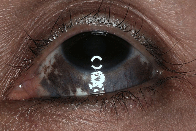
[Image credit: "Nevus of Ota" by Brian Boxer Wachler is licensed under CC BY-SA 4.0 International license.]
Hyperpigmentation can appear during puberty and adulthood in people who have nevus of Ota, even if it wasn’t present at birth. The lesions’ color ranges from brown to gray-blue to gray-black. More than 66% of patients with nevus of Ota have hyperpigmentation on the white of one of their eyes (the sclera). This is associated with an increased risk of glaucoma development.
Nevus of Ota usually affects one side of the face. The color depth of the lesion may appear different with changes in weather, fatigue or menstrual cycle. It is also common for the color to darken after puberty.
Causes
Nevus of Ota is most common in people of Asian and African descent. White people with nevus of Ota are at higher risk of developing malignant melanoma, however. Nevus of Ota affects women at a 5:1 ratio compared to men.
Researchers have yet to find a precise cause for nevus of Ota, but theories of possible causes include:
Genetic mutations
Hormonal influence
Exposure to radiation
Similar conditions
Conditions similar to nevus of Ota include:
Nevus of Ito – Nevus of Ito presents as hyperpigmented lesions on the upper arm or shoulder. The lesions have the same range of color as nevus of Ota.
Congenital melanocytosis (Mongolian spots) – This condition is also called dermal melanocytosis. Mongolian spots are birthmarks often distributed along the lower back. They range in color from blue-green to black and vary in shape. Like nevus of Ota, Mongolian spots are most common in people of Asian or African descent. These spots usually disappear by age two, but can last into adulthood. Occasionally, Mongolian spots appear on the mouth or temple.
Drug-induced hyperpigmentation – Certain drugs can cause hyperpigmentation of the skin, which usually fades after treatment ends.
Melasma – Melasma is a skin condition that can cause blotches or spots to appear on the face. Most of the time, these marks are slightly darker than the person’s skin color. They can look bluish-gray in people with darker skin.
Nevus of Hori – Nevus of Hori often appears at around 30 to 50 years old, and is most common in women of Asian descent. It usually presents as blue-brown patches on both sides of the face, but does not affect the eyes.
READ NEXT: What Is Ochronosis
Potential complications
Although it is unlikely, nevus of Ota can lead to complications. People with hyperpigmentation in the sclera are at a higher risk of developing glaucoma. About one in 400 individuals with nevus of Ota affecting the eye develop choroidal melanoma, a type of uveal melanoma.
Uveal melanoma is rare, but it is also the most common ocular melanoma (eye cancer) in adults that occurs inside the eye. Symptoms vary based on the location of the tumor, but the risk of metastasis (spreading to other body parts) exists regardless of its location within the uveal tract. It is most common in Caucasian adults in their 50s and 60s.
Uveal melanoma is serious, and early detection and treatment can be life-saving. You should contact an eye doctor if you notice any of the following symptoms or signs:
A change in vision
Drifting spots in your field of vision
Flashes of light
A dark spot on the iris
A change in pupil shape or size
The eyeball has shifted in its socket
A condition outside the eye can sometimes increase the risk of developing cancer inside the eye. Because you may not be able to see the signs and symptoms, it is important to get yearly comprehensive eye exams.
Treatment
Treatment for nevus of Ota can be divided into eye-related and skin-related:
Eye-related treatment
A doctor will perform a comprehensive eye exam in order to determine the extent that nevus of Ota is involved with the eye. Glaucoma screening every year is recommended. They may also check for any uveal melanomas. If uveal melanoma is found, management will depend on its size and location.
The following procedures will likely be involved:
Surgical resection – The tumor is removed from the eye by a vitreoretinal surgeon.
Radiotherapy – Radiation is applied to kill the cancerous cells.
Transpupillary thermotherapy – A laser treatment that uses infrared light to kill the tumor.
Enucleation – The eye is removed and replaced with an artificial eyeball. This is usually performed when the tumor has grown too large for other treatments to be effective.
Skin-related treatment
Laser surgery is often the primary cosmetic treatment for nevus of Ota. It will likely have to be repeated multiple times for the hyperpigmentation to be reduced or removed.
Some nonsurgical methods of treatment are:
Chemical peeling – A chemical peel removes layers of skin and reveals the younger skin underneath.
Dermabrasion – The top layers of skin are “sanded” away to reveal the younger layer underneath.
Cryotherapy – Extreme cold is applied to the affected parts of the skin to kill the tissue there.
When to see a doctor
You should talk with your eye doctor if you think you have nevus of Ota, even if you don’t notice any symptoms that indicate potential complications. That way, routine checkups can be scheduled to help prevent any complications from the condition.
SEE RELATED: Linear Nevus Sebaceous Syndrome
Nevus of Ota and Ito. StatPearls. March 2022.
Nevus of Ito. GARD. November 2021.
The Treatment of Hori's Nevus by New Combination Treatment without Side Effects: Dr. Hoon Hur's Golden Parameter Therapy and Dr. Hoon Hur's Optimal Melanocytic Suicide-2 Parameter Therapy. Symbiosis Online Publishing. December 2017.
Oculodermal melanocytosis (nevus of Ota). Eyewiki. January 2022.
Mongolian spots: How important are they? World Journal of Clinical Cases. November 2013.
Drug induced pigmentation. StatPearls. July 2022
Melasma: signs and symptoms. American Academy of Dermatology Association. February 2022.
Novel treatment of Hori's nevus: A combination of fractional nonablative 2,940-nm Er:YAG and low-fluence 1,064-nm Q-switched Nd:YAG laser. Journal of Cutaneous and Aesthetic Surgery. December 2015.
Uveal melanoma. American Academy of Ophthalmology. September 2021.
Intraocular (Uveal) Melanoma Treatment (PDQ®)–Patient Version. National Cancer Institute. July 2021.
Treatment of uveal melanoma: where are we now? February 2018.
Chemical peels. Cleveland Clinic. March 2021
Dermabrasion. Cleveland Clinic. September 2020
Cryotherapy. Cleveland Clinic. May 2020
Page published on Wednesday, November 16, 2022
Medically reviewed on Wednesday, October 26, 2022


