Causes and symptoms of optic atrophy

What is optic nerve atrophy?
The optic nerve is made of over one million nerve fibers that send light signals from the retina to the brain. Optic nerve atrophy is the death of a portion of these fibers, leading to blurry or dim vision, side vision loss and altered color vision. Causes include injury, inflammation and pressure.
The retina is the light sensitive tissue at the back of the eye. It is made of many layers of nerve cells. Photoreceptor cells are cells in the retina that convert light into electrical signals.
Electrical signals are sent from photoreceptor cells to retinal ganglion cells, the nerve cells that relay these signals to the brain. This is done via thin, long fibers, called axons, that extend from the retinal ganglion cells and eventually come together in a bundle to help form the optic nerve.
This nerve is also known as Cranial Nerve II. It leaves the retina and travels to a part of the brain called the occipital lobe, where vision is processed. The optic nerve can be seen in the center of the retina as a pinkish circle about 1.5 mm in diameter.
What are symptoms of optic nerve atrophy?
Vision loss can range in severity and type. Damage to the optic nerve can affect central vision, side vision and color vision.
When the ability of the optic nerve to transfer signals to the brain is interrupted, an individual may experience the following:
Dimming of vision in affected eye
In children, it may cause uncontrolled up and down or side to side eye movements called nystagmus.
If any of these symptoms are noticed, it is critical to contact an eye doctor.
Causes and types of optic nerve atrophy
Optic nerve atrophy is caused by death of retinal ganglion cell axons that make up the optic nerve. Once optic nerve fibers are lost, they cannot regenerate. Optic atrophy is considered to be the end stage of the underlying disease.
The most common cause of optic nerve atrophy is poor blood flow, also known as “ischemia.” This is most often seen in older adults. Glaucoma is another common condition that can lead to optic nerve fiber loss.
Optic nerve atrophy is often categorized by its underlying cause, which can include a large number of conditions:
Due to damage to the optic nerve inside the eye:

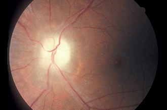
Optic nerve atrophy from untreated raised intraocular pressure. [Image credit: "Optic atrophy" by International Centre for Eye Health is licensed under CC BY-NC 2.0]
Ischemia – decreased blood supply due to central retinal artery occlusion or carotid artery occlusion
High intraocular pressure – high pressure in the eye, often due to glaucoma
Optic neuritis – inflammation of the optic nerve
Hypoxia – decreased oxygen supply
Conditions causing papilledema – swelling of the optic nerve
Orbital cellulitis – infection of the eyeball and tissues surrounding the eyeball, usually occurring in young children
Coats' Disease – an abnormality of the eye’s blood vessels. This rare condition manifests as enlarged, twisted veins that may leak, limiting blood flow and oxygen to the retina.
Due to central nervous system (brain and spinal cord) diseases:
Inflammatory diseases – such as multiple sclerosis, meningitis
Ischemia – decreased blood supply due to conditions such as temporal arteritis
Stroke – usually due to decreased blood flow to the brain
Due to damage along the optic nerve pathway from the eye to the brain:
Tumors – that compress the nerve
Hydrocephalus – the build-up of fluid in the cavities of the brain
Injury – due to head trauma or injury that penetrates the optic nerve
Neurodegenerative disorders – such as Alzheimer’s and Parkinson’s disease
READ MORE: Idiopathic Intracranial Hypertension (IIH)
Due to toxins or lack of nutrition:
Toxicity – such as methanol poisoning from home-brewed alcohol
Vitamin deficiencies – a rare complication causing optic atrophy in both eyes due to deficiency of vitamin B complex (typically occurs during famine and war)
Due to abnormal fetal development:
Improperly developed optic nerve – from optic nerve hypoplasia (underdevelopment of the optic nerve)
Autosomal dominant optic atrophy – degeneration of the optic nerve, often beginning in childhood.
Pfeiffer syndrome – a rare genetic disorder that affects the skull, face and eyes.
Sometimes even after evaluation for all of these conditions, no specific cause is able to be identified.
READ MORE: A guide for parents of visually impaired children
How is optic atrophy diagnosed?
An eye doctor will note a pale appearance of the optic nerve when looking inside the eye during a dilated eye exam. This appearance is usually present about four to six weeks after damage to the optic nerve. A comprehensive eye exam will be required to diagnose optic atrophy and determine the underlying cause. The exam may include:
Dilated exam – nerve fiber loss causes the optic nerve to look pale
Medical history
Peripheral (side) vision assessment
Pupil reaction
Eye alignment
Head position
Presence of nystagmus
Additional testing that may be required:
Brain imaging such as MRI scan
Electroretinography (ERG)
Visual evoked potential (VEP)
Blood tests
Routine, dilated eye exams are the best strategy for early detection of eye conditions such as optic nerve atrophy. During a dilated eye exam an eye doctor can notice early changes in the back of the eye that can indicate damage to the optic nerve. If signs of optic nerve damage are present, further evaluation and additional testing may be needed in order to treat the underlying cause.
Can optic nerve atrophy be reversed?
Optic atrophy cannot be reversed. But, managing blood pressure and maintaining a heart healthy lifestyle can help to decrease the risk of many eye conditions, including optic atrophy.
In addition, it is important to routinely wear eye and head protection as well as car seat belts to prevent eye injuries. If you have been diagnosed with glaucoma, it is critical to follow the management plan prescribed by your doctor.
READ MORE: Eye Safety Basics
Are there any treatments for optic nerve atrophy?
Once they are lost, optic nerve fibers do not recover. Early diagnosis and treatment of the underlying causes of optic atrophy are important in preventing further loss of vision.
If permanent vision impairment has occurred due to optic nerve atrophy, daily activities can become a challenge. Low vision eye doctors are trained to perform a specialized eye exam.
These doctors can assist those who have suffered a loss of functional vision. They are knowledgeable about the latest devices and technology aids for those affected by vision impairment. This includes magnifiers, special electronic devices and other assistive technology.
Optic atrophy. American Academy of Ophthalmology. EyeWiki. January 2022.
Photoreceptors. Biomechatronics. 2019.
Optic nerve. Ocular Pathology. 2020.
Optic nerve atrophy . American Association for Pediatric Ophthalmology and Strabismus. March 2020.
Optic atrophy. Kellogg Eye Center Michigan Medicine. Accessed March 2022.
Periorbital and orbital cellulitis. Journal of the American Medical Association. January 2020.
Types of stroke. Centers for Disease Control and Prevention. August 2021.
Hydrocephalus. American Association of Neurological Surgeons. Accessed March 2022.
Alzheimer’s disease, dementia and the eye. American Academy of Ophthalmology. May 2019.
Optic neuropathies: the tip of the neurodegeneration iceberg. Human Molecular Genetics. October 2017.
Optic nerve hypoplasia. National Organization for Rare Disorders (NORD). Accessed March 2022.
Dominant optic atrophy. Genetic and Rare Diseases Information Center. December 2015.
Optic nerve atrophy. MedlinePlus Medical Encyclopedia. August 2020.
Optic atrophy. StatPearls — NCBI Bookshelf. February 2022.
Nutritional optic neuropathy. American Academy of Ophthalmology. EyeWiki. February 2022.
Page published on Wednesday, April 27, 2022


