Hypertelorism: Definition, causes, diagnosis and management

What is hypertelorism?
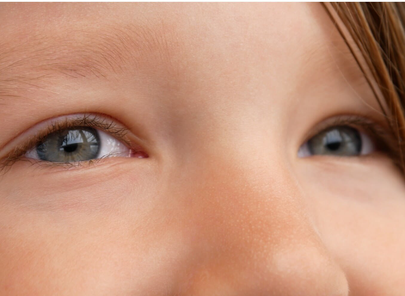
Hypertelorism is a medical term for having widely spaced eyes that are often caused by birth defects and genetic disorders. Hypertelorism isn’t a diagnosis or syndrome in and of itself. Instead, it is often a physical finding in many craniofacial syndromes.
Hypertelorism develops before birth. It is due to an alteration in facial development during weeks 4-8 of development during pregnancy. Some sources estimate hypertelorism occurs in one in 20,000 births.
What causes hypertelorism?
Hypertelorism is thought to be caused by an alteration in an embryo’s facial development during the first three months of pregnancy. This alteration prevents the movement of the orbits (bony eye sockets) toward the middle of the face.
These alterations can be caused by mechanic factors, such as:
Abnormal development of the forehead bones and the base of the skull.
Premature fusion of the bony plates of the skull.
A mass pushing the bones apart.
Hypertelorism can also be associated with craniosynostosis syndromes. Craniosynostosis is the premature closure of certain bones in the skull while brain growth continues at a normal pace. This results in a misshaped appearance of the skull. Craniosynostosis can cause hypertelorism. Craniosynostosis may exist in isolation or with syndromes such as:
Crouzon syndrome
Pfeiffer Syndrome
Diagnosing hypertelorism
Hypertelorism is usually noticed at birth. It may be observed prior to birth with a fetal ultrasound. There are so many possible underlying factors for hypertelorism. Therefore, evaluation requires specific and precise measurements.
When hypertelorism is identified, a complete physical examination is required to identify any congenital abnormalities.
The initial diagnosis is usually made in a clinical assessment. Further evaluation can be done with imaging such as a CT scan or an MRI.
There are three facial measurements needed to determine if a person has hypertelorism:
Inner canthal distance (ICD) – the distance (in mm) between the nasal/inner corners of the two eyes
Outer canthal distance (OCD) – the distance (in mm) between the temporal/outer corners of the two eyes
Interpupillary distance (IPD) – the distance (in mm) between the center of the two pupils
If all three of these measurements are above the 95th percentile of that expected in an average person, orbital hypertelorism is present. This is also known as true hypertelorism.
Telecanthus (pseudo-hypertelorism) occurs when the ICD is higher than average, but the OCD and IPD are not. Disorders that often feature telecanthus include:
Down syndrome
Fetal alcohol syndrome
Ehlers-Danlos syndrome
Klinefelter syndrome
During an evaluation of hypertelorism, a doctor may also order a dysmorphology exam. This exam is completed on newborns and focuses on case history and physical examination. It looks to identify factors that may lead to a syndrome.
The findings of this exam often determine underlying causes of a person’s hypertelorism or telecanthus.
Management and treatment
Many cases of hypertelorism can be monitored and do not need treatment. Surgery is a treatment for hypertelorism that addresses the cosmetic issue. The goal of this surgery is to bring the two orbits closer together.
This surgical procedure is complex and requires a team of craniofacial surgeons and ophthalmologists. It is also essential for the surgeons to know the underlying cause of hypertelorism. This helps them determine if a surgical correction is appropriate.
The timing of the surgery to treat hypertelorism is important. The ideal time period is between 5 and 7 years old. Prior to 5 years old, the bones may not be strong enough to support a positive surgical outcome. After the age of 7, there is an increased concern of a negative effect on the child’s self-esteem.
Possible surgeries for hypertelorism:
Box osteotomy – A piece of bone is removed from between the eyes. The bones containing the eye sockets are then moved to the space opened by the removed bone.
Facial bipartition – The nasal ridge (area from the tip of the nose to its root) is removed from between the eyes. Both halves of the upper face are then rotated to bring the eyes closer together.
Box osteotomy and facial bipartition are the two main surgeries for hypertelorism. A procedure is chosen based on an individual’s facial bone structure. The degree of hypertelorism and the presence of any associated conditions are also considered.
Hypertelorism is not a disease. But it is often associated with congenital issues and craniofacial synostosis. Visual issues resulting from hypertelorism are rare. Patients with hypertelorism should have a full comprehensive eye examination in order to best understand and (when necessary) manage the condition.
Pediatric hypertelorism. Children’s Health. Accessed December 2022.
Hypertelorism. StatPearls. July 2022.
Hypertelorism. EyeWiki. American Academy of Ophthalmology. September 2022.
Hypertelorism. Dell Children’s Craniofacial and Pediatric Plastic Surgery Program. Accessed December 2022.
Syndromic craniosynostosis. Children’s Hospital of Philadelphia. Accessed December 2022.
Aarskog syndrome. Genetic and Rare Diseases Information Center. November 2021.
Fetal alcohol spectrum disorders. Centers for Disease Control and Prevention. November 2022.
Anthropometric measurement. StatPearls. September 2022.
Orbital hypertelorism. Children’s hospital of Philadelphia. March 2014.
Page published on Thursday, December 15, 2022
Page updated on Tuesday, January 10, 2023
Medically reviewed on Friday, November 18, 2022


