How sickle cell disease affects vision

What is sickle cell disease?
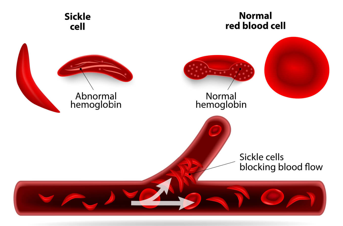
Sickle cell disease (SCD) is a disorder of red blood cells. Healthy cells have a flexible disc shape that lets them move easily through blood vessels. People with SCD have red blood cells that are sticky and shaped like a sickle. This causes them to stick and block blood flow to the body and eyes.
Red blood cells contain a protein called hemoglobin. Hemoglobin helps deliver oxygen to the tissues of the body. In sickle cell disease, this protein — called hemoglobin S — is abnormal. This causes the red blood cells to have a distorted “C” shape. These cells are stiffer and tend to clump together, blocking blood flow in blood vessels and depriving the body’s tissues of oxygen and nutrients. This can lead to symptoms such as anemia and periods of pain and result in organ damage.
Sickle cell retinopathy is the most common cause of vision loss in people with SCD. Signs and symptoms of sickle cell disease begin in early childhood, sometimes before a child’s first birthday. Because SCD can lead to serious medical and visual complications, all 50 states require that newborns are screened for this disease. This condition affects millions worldwide and is most commonly seen in people of African, Hispanic and Caribbean descent.
Symptoms
Sickled red blood cells can block blood flow to different parts of the body, including the eye. The retina is the most common part of the eye that is affected. But SCD can also damage both the front and the back of the eye, leading to severe complications.
Sickle cell retinopathy
Sickle cell retinopathy is caused by changes to the blood vessels of the retina due to blood flow blockage. It can occur in one or both eyes and is the most common cause of vision loss in people with SCD. It can affect up to 42% of people with SCD beginning in their teen years.
Pressure buildup can cause blocked blood vessels to burst and bleed, damaging the retina and causing vision loss. Symptoms of sickle cell retinopathy include:
Sudden floaters
Light flashes
Dark curtain closing over vision
Side vision loss
There are two types of sickle cell retinopathy. Non-proliferative sickle cell retinopathy (NPSR) is the less serious type. It is not as likely to cause vision loss. Proliferative sickle cell retinopathy (PSR) is more likely to lead to vision loss, affecting 10% to 20% of people.
Proliferative sickle cell retinopathy is the formation of abnormal, leaky blood vessels on the inner surface of the retina or vitreous. It increases the risk of retinal detachment or vitreous hemorrhage (see below).
During a dilated eye exam, an eye doctor examining the retina will be able to see these burst blood vessels. They may also note a typical “sea fan” pattern on the retina, indicating PSR.
Sickle cell maculopathy
The macula is the small central part of the retina responsible for detailed, central vision. Decreased blood flow to this part of the retina can lead to damage and thinning of the tissue. This results in decreased and distorted central vision as well as blind spots.
Vitreous hemorrhage
Proliferative sickle cell retinopathy can cause a vitreous hemorrhage. A vitreous hemorrhage is bleeding in the vitreous humor — the gel-like fluid that helps maintain the eye’s shape — in the center portion of the eye. This can occur when new, leaky abnormal blood vessels burst and leak blood inside the eye.
Retinal detachment
Retinal detachments occur when the retina pulls away from the layer of blood vessels that nourishes it. This can lead to severe vision loss. Retinal detachments are more likely to occur in parts of the retina that have tears due to poor blood flow or traction caused by abnormal blood vessels and scar tissue.
Hyphema
An eye injury can cause hyphema, the presence of blood in the anterior chamber. This is the space between the cornea — the clear dome-shaped tissue at the front of the eye — and the iris — the colored part of the eye.
Glaucoma can result from hyphema, although this is rare. Glaucoma results when increased pressure inside the eye causes damage to the optic nerve. This is the nerve that carries signals from the eye to the brain.
Increased pressure in the eye can also increase the risk of retinal artery occlusion — the blockage of an artery in the retina. This can damage the retina and cause vision loss.
It is very important to contact an eye doctor if you have SCD or sickle cell trait (SCT) and experience an eye injury.
Additional complications
People with SCD may experience pain and swelling of the lacrimal gland. The lacrimal gland is located above the eye and is responsible for tear production.
The blood vessels of the conjunctiva — the clear tissue covering the eye — can take on a “comma-shape” in people with SCD. This does not affect vision but may indicate the level of damage to the body’s blood vessels.
The cornea may become cloudy in people with SCD due to injury, inflammation or infection. Depending on the location, this can lead to blurred vision.
Causes
Sickle cell disease is a genetic condition. A copy of the hemoglobin S gene must be passed down from both parents for the disease to be inherited.
There are a few different types of sickle cell disease, and some are quite rare. The most common types include:
HbSS – The most severe type of SCD in which an individual has inherited the hemoglobin S gene from both parents. Vision loss is less common as the retina is less affected by this type.
HbSC – A less severe form of SCD in which an individual inherits an atypical hemoglobin “C” gene from one parent and hemoglobin S from the other parent. Vision loss is more common as the retina is more affected by this type.
HbS beta thalassemia – A type of SCD in which an individual inherits a gene mutation for beta thalassemia – a blood condition that decreases the production of hemoglobin – from one parent and hemoglobin S from the other parent. The severity of SCD varies with the type of gene inherited. Eye symptoms are not usually present.
Some people inherit a normal hemoglobin gene — called hemoglobin A — from one parent and a hemoglobin S gene from the other parent. These people are considered to have sickle cell trait.
People with SCT generally do not have health problems. But they may (rarely) exhibit symptoms if their body experiences stress, such as dehydration or severe exhaustion. Individuals with SCT can pass down the gene to their children.
Risks and complications
The most common complications of SCD are episodes of severe pain. Another complication is the increased risk of organ damage because of inadequate blood flow to the tissues. Individuals with SCD can begin to show signs and symptoms of the condition in very early childhood.
The most common symptoms include:
Anemia – A condition in which there are too few red blood cells to carry oxygen to the body’s tissues. The red blood cells break down more easily in SCD and do not last as long, leading to anemia. This can cause fatigue, dizziness and breathing difficulties. In children, it can result in delays in development and growth.
Jaundice – A condition that gives a yellow color to the eyes and skin. This is due to the byproducts of the fast rate of red blood cell breakdown.
Periods of pain – Due to blockage of blood flow because the abnormal red blood cells have clumped together, starving the tissue.
Acute chest syndrome – A condition that causes breathing difficulty and low levels of oxygen in the body. It can injure the lungs and is a medical emergency.
Recurrent infections – Damage to the body’s organs results in decreased immunity. Pneumonia, flu and meningitis are more likely in people with SCD and may be life-threatening.
Pulmonary hypertension – A condition that typically affects adults and can lead to heart failure due to high blood pressure in the arteries of the lungs.
Avascular necrosis – A condition that decreases blood flow to the bones, damaging bone tissue and causing joint pain. The hip joint is a common location.
Blood clots – A clot in a deep vein caused by the clumping of abnormal red blood cells.
Stroke – A blood clot that has blocked blood flow to the brain.
Diagnosis
Early indications of eye damage can be seen during a dilated eye exam, even before vision is affected. Due to this, it is important for people with SCD to get comprehensive eye exams on a routine basis to help prevent vision loss.
An eye doctor may order additional tests to manage the condition, including:
Fluorescein angiography – This procedure requires fluorescein dye to be injected into the arm, which travels to the eye’s blood vessels. The dye lights up areas with choroidal neovascularization. This allows the doctor to identify where leaking blood vessels in the retina are located.
Optical coherence tomography (OCT) – This procedure provides 3D images of cross-sections of the retina, indicating the presence of retinal thinning in SCD. It is painless and does not require an injection.
Optical coherence tomography angiography – This procedure shows whether the blood vessels of the retina and choroid are blocked. It can also detect the presence of new abnormal blood vessels.
Treatment and prognosis
If small areas of abnormal blood vessels are present in the retina, an eye doctor will closely monitor them rather than begin treatment. They do this because these areas often go away on their own.
If an eye doctor diagnoses proliferative sickle cell retinopathy, the first line of treatment is usually retinal laser photocoagulation to prevent retinal detachment or bleeding in the vitreous gel. The goal is to slow the loss of vision and potentially improve vision that has worsened.
Retinal laser photocoagulation
Laser photocoagulation uses a laser to generate heat and create a scar that can seal off abnormal and leaky blood vessels. This scar can also weld down small retinal detachments, walling them off so they do not progress. It is an outpatient procedure and is relatively painless.
Anti-VEGF therapy
Doctors may also inject anti-VEGF drugs, such as the medication bevacizumab (brand name Avastin), into the eye. They do this to block the excessive production of VEGF — vascular endothelial growth factor. In normal amounts, VEGF helps to maintain the retina.
But excess VEGF stimulates choroidal neovascularization. This is the growth of new, abnormal and leaky blood vessels in the choroid — the layer under the retina. Multiple anti-VEGF injections are often required.
Surgery
Surgery is usually saved for cases in which proliferative sickle cell retinopathy has become advanced. If bleeding in the vitreous hasn’t cleared or there is a retinal detachment or other complications, surgery may be required. However, this carries a higher risk.
Systemic treatments
Red blood cell exchange transfusions and the drug hydroxyurea (brand name Hydrea) may be used to manage sickle cell retinopathy. The drug reduces the amount of abnormal sickle hemoglobin (HbS) circulating in the body. This helps to increase blood flow to the tissues of the body, including the eye.
Two medications have also recently become available. Voxelotor is a drug that helps prevent red blood cells from sickling and breaking down. Crizanlizumab helps to prevent the blockage of blood vessels.
Stem cell therapy from a sibling has been found to be effective as well.
Prognosis
The severity of sickle cell disease varies with each individual. So, the prognosis depends on many different factors, such as the type of disease and early intervention. Vision loss is less common in people with HbSS type and more common in those with HbSC type.
When to see a doctor
Yearly comprehensive eye exams are important for people with sickle cell disease. People who have proliferative sickle cell retinopathy will need to be monitored more frequently.
A team of healthcare professionals that are trained to manage sickle cell disease is critical for preventing short-term and long-term medical and vision issues.
People with SCD need to take certain precautions. Avoiding decongestants is recommended. Decongestants constrict blood vessels and can increase the risk of blood vessel blockage.
Experts also recommend that people with SCD avoid exhaustion, strenuous exercise or labor, high altitudes, cold weather, and swimming in cold water. Preventing infections by frequent hand washing, vaccinations, avoiding sick people and regular medical and dental check-ups is also advised.
In addition, experts recommend that individuals with sickle cell disease stay hydrated with eight to 10 glasses of water daily. Folic acid (folate) supplements can help to decrease the risk of severe anemia. Eating a diet rich in fruits, vegetables and whole grains is important for increasing immunity and staying healthy.
Finally, it is important for individuals with SCD to always be prepared with medications and instructions from their doctor in case of an emergency.
Sickle cell disease. MedlinePlus Genetics. July 2020.
What is sickle cell disease? Centers for Disease Control and Prevention. August 2022.
Sickle beta thalassemia. National Organization for Rare Disorders. Accessed May 2023.
Sickle cell retinopathy. EyeWiki. American Academy of Ophthalmology. December 2022.
Retinopathy and sickle cell disease. St. Jude Children’s Research Hospital. September 2022.
Sickle cell retinopathy: An update on management. Retina Specialist. December 2022.
Sickle cell retinopathy. The American Society of Retina Specialists. Accessed May 2023.
Eye problems and sickle cell trait: Learn how you can help protect your vision. Centers for Disease Control and Prevention. Accessed May 2023.
Ocular complications in sickle cell disease: A neglected issue. Journal of Ophthalmology. August 2020.
Sickle cell disease. Johns Hopkins Medicine. Accessed May 2023.
Complications of sickle cell disease. Centers for Disease Control and Prevention. May 2022.
Pathophysiological role of VEGF on retinal edema and nonperfused areas in mouse eyes with retinal vein occlusion. Investigative Ophthalmology and Visual Science. September 2018.
Page published on Tuesday, May 16, 2023
Page updated on Tuesday, May 23, 2023


