What is corneal topography, and why is it important?

Corneal topography (also known as computerized corneal mapping or computer-assisted videokeratoscopy) is a diagnostic tool that creates a color-coded, three-dimensional map of the surface of the cornea — the eye’s outermost layer.
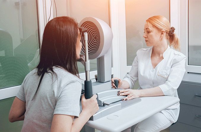
The technique is performed with an instrument called a corneal topographer, which projects rings of light onto the corneas and then analyzes the reflected images using special technology.
This process is helpful for ophthalmologists to diagnose, monitor and treat specific eye conditions that affect the cornea. It is also used for things like contact lens fittings and eye surgery preparation.
Corneal topography is not a part of every comprehensive eye exam, but if you or your ophthalmologist have concerns about your cornea health, it may be recommended.
What is corneal topography used for?
A healthy, normal cornea has even curvatures and a smooth surface. A corneal topography scan provides insight into any curvature or surface abnormalities that may be present. Such abnormalities can indicate diseases, scarring and other conditions that may affect the health of your cornea.
Corneal topography is used for several reasons, including the following:
Diagnosing, monitoring and treating eye conditions involving the cornea.
Evaluating corneal injuries or deformities.
Preparing for eye surgery and monitoring postoperative ocular health.
Measuring corneal curvature (for astigmatism) and corneal depth.
Evaluating the corneal nerve.
Fitting for special contact lenses.
Contact lens fitting via corneal topography is recommended for people who need contact lenses after a refractive surgery, as well as those who have astigmatism or keratoconus — a progressive eye disease that causes the cornea to thin and bulge into a cone-like shape.
Ultimately, a hard contact lens, such as a gas permeable (GP) lens, is advised for such cases.
SEE RELATED: Answers from an eye doctor about keratoconus
Conditions identified using corneal topography
What specific conditions are monitored with corneal topography? Ophthalmologists may recommend an exam to detect and/or monitor the following:
Corneal growths such as pterygium (surfer’s eye).
Corneal abrasions and ulcers.
Corneal trauma, or scarring near the cornea that can result in a change in shape.
Astigmatism, especially after corneal transplant surgery (keratoplasty).
Keratoconus (detecting early signs of the disease and monitoring progression).
If you have an eye disease that requires steady monitoring, your eye doctor will let you know how frequently you’ll need a corneal topography exam.
Corneal topography is used for surgical planning
Surgical procedures require detailed planning, especially those that involve the cornea. Specific surgeries that make use of corneal topography include those designed to reshape — and even replace — the cornea.
Following a corneal transplant, an eye surgeon may use corneal topography to identify when and where stitches should be removed.
Corneal crosslinking is a procedure performed to strengthen the corneal tissue of people who have keratoconus. In these cases, corneal topography scans are also used postoperatively to monitor the eye.
In refractive surgeries such as LASIK, the cornea is reshaped to correct refractive errors such as myopia (nearsightedness). Corneal topography is used to direct an eye surgeon exactly how and where to reshape the cornea.
Cataract surgery involves replacing the eye’s natural lens with an intraocular lens (IOL). A corneal topography scan can help determine the best IOL to be inserted during this procedure.
What to expect during a corneal topography exam
Corneal topography is quick, painless and noninvasive. The process takes just a few seconds, though it may be repeated several times for accuracy. Contact lenses should be removed before a scan.
During a corneal topography scan, you will be seated in front of a large, bowl-like instrument (called a corneal topographer) with lighted circles inside. This structure has a forehead and chin rest, which are used to keep your head and eyes aligned during the scan.
Light is then projected onto your corneas, and several pictures are taken as you look into the corneal topographer. Your eye doctor will review these pictures with you either during your appointment or at a follow-up appointment, then recommend next steps for your condition based on their findings.
Although corneal topography is not recommended for every single patient, it is still important for everyone to undergo a comprehensive eye exam once a year. This is the best way to maintain good eye health and detect eye problems big and small.
READ NEXT: Eye exams: 5 reasons why they are important
Page published on Wednesday, March 10, 2021


